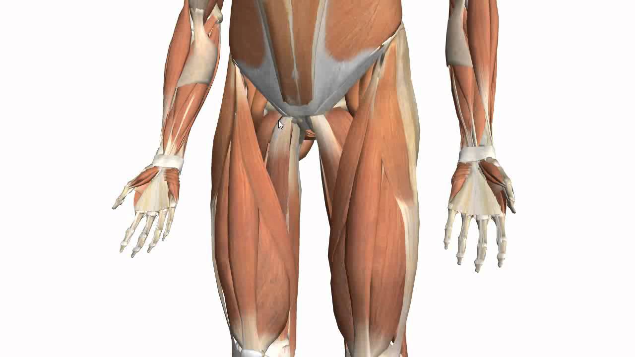
The tendons for these muscles begin at your ischial tuberosity, or ischium (the. Use the mouse scroll wheel to move the images up and down alternatively use the tiny arrows (>>) on both side of the image to move the images. .16 penile numbness and perineum tenderness.18 any suggested exercises or stretches?.22 leg musculature 209 elbow tendonitis and saddle sores.
Here some resume from the keyword to help you find your search, the owner of the copyright is the original owner, I hope this will help you a lot
.16 penile numbness and perineum tenderness.18 any suggested exercises or stretches?.22 leg musculature 209 elbow tendonitis and saddle sores. Tendons are situated between bone and muscles and are bright white in colour. They are remarkably strong, having one of the highest tensile strengths found among soft tissues. The thigh bone, or femur, is the large upper leg bone that connects the lower leg bones (knee joint) to the pelvic bone (hip joint). Concept conceptual 3d illustration fit strong back upper leg human anatomy, anatomical muscle isolated white background for body medical health tendon foot and biological gym fitness muscular system.
Tennis Leg and Achilles Tendonitis: Confusing The Two Can ... Visit full article here : Bing Choose from 500 different sets of flashcards about anatomy muscle anatomy_ upper leg on quizlet. Lateral (fibular) collateral ligament (fcl) upper part middle part lower part popliteus tendon (pt) upper part i. The lower leg is comprised of two bones, the tibia and the smaller fibula. Muscle/tendon inflammation and pain along anterio… The peroneus longus tendon moves out of place behind the lateral malleolus of your ankle and then snaps back into.
Palmar region , arteries (illustrations:
Collectively, they act to dorsiflex and invert the foot at the ankle joint. The lower leg is comprised of two bones, the tibia and the smaller fibula. The thigh bone, or femur, is the large upper leg bone that connects the lower leg bones (knee joint) to the pelvic bone (hip joint). This may result in tendon subluxation; What are the functions of patella.
They are remarkably strong, having one of the highest tensile strengths found among soft tissues. Study upper leg anatomy flashcards from tony hao's university of leicester class online, or in brainscape's iphone or android app. Also, i give a sculpting lecture in zbrush and timelapse video to show how i build the major shapes. Muscle/tendon inflammation and pain along anterio… How does achilles tendon rupture occur… why are achilles piercings dangerous?
Muscles of the Thigh Part 2 - Medial Compartment - Anatomy ... Visit full article here : Bing Palmar region , arteries (illustrations: Current techniques have tended to anatomical reconstruction of the lcl, pt and pf. Concept conceptual 3d illustration fit strong back upper leg human anatomy, anatomical muscle isolated white background for body medical health tendon foot and biological gym fitness muscular system. Lie prone on a hamstring curl machine. The tendons of the edl can be palpated on the dorsal surface of the foot.
Use the mouse scroll wheel to move the images up and down alternatively use the tiny arrows (>>) on both side of the image to move the images.
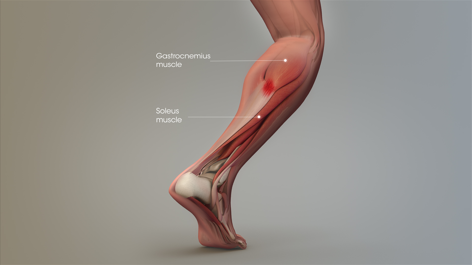
1280 x 1520 jpeg 166 кб. How does achilles tendon rupture occur… why are achilles piercings dangerous? Lateral (fibular) collateral ligament (fcl) upper part middle part lower part popliteus tendon (pt) upper part i. This article will discuss the anatomy and function of the achilles tendon. Common tendon of superficial posterior leg muscles;
The pads of the machine are situated at the achilles tendon. An anatomical and biomechanical study. The tendons of the edl can be palpated on the dorsal surface of the foot. Tendon, tissue that attaches a muscle to other body parts, usually bones. Plantar flexion of the foot, ankle joint stabilizer.
29 Best Muscle labeling "PTA" images | Muscle, Muscle ... Visit full article here : Bing Superficial veins of upper limb , anatomy : The calcaneal tendon, also known as the tendon of achilles, is a posterior leg tendon — a fibrous connective tissue that joins muscles in the back of the leg. Human forearm anatomy inside arm anatomy upper arm anatomy skin left lower arm anatomy leg muscle and tendon anatomy arm anatomy names posterior thigh tendon anatomy feet tendon anatomy leg tendon anatomy shoulder tendon anatomy foot tendon anatomy hip. Collectively, they act to dorsiflex and invert the foot at the ankle joint. The tendons that control movement in your hands, wrists and fingers run through your forearm.
Hands are outstretched, holding onto the handles of the bench.
The large achilles tendon is the most important tendon for walking, running, and jumping. Use the mouse scroll wheel to move the images up and down alternatively use the tiny arrows (>>) on both side of the image to move the images. Tendons are fibrous cords attached to muscles and bone. An anatomical and biomechanical study. Superficial veins of upper limb , anatomy :


Posting Komentar untuk "Upper Leg Tendon Anatomy"